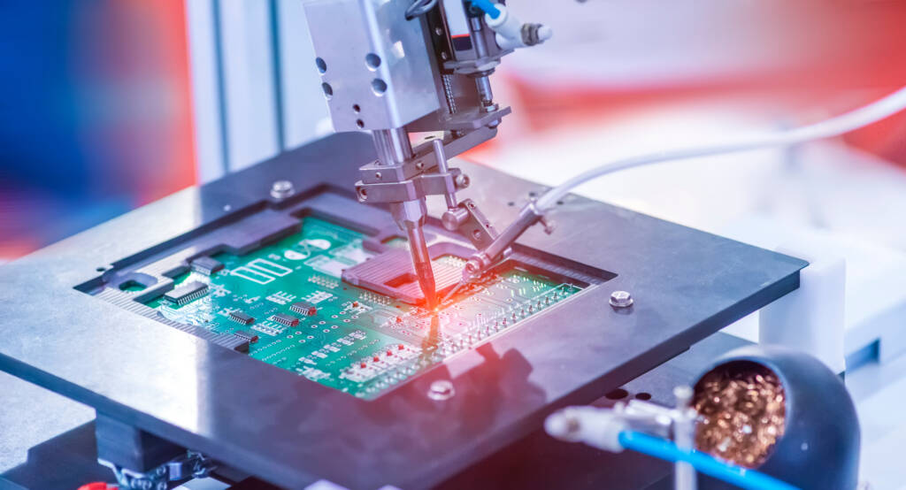
Scanning Electron Microscopy (SEM) is a technique used to characterize cross sections of solder joints to identify a root cause to a soldering failure. Although there are many different detection methods that can be used with a SEM, the two most common detection methods are Backscattered Electron (BSE) detection and Secondary Electron (SEI) detection.
Many times, BSE and SEI are erroneously used interchangeably. It is important to remember that these two detection methods produce micrographs that are better used to observe two different features of a material. SEI micrographs are better used for looking at surface features of a material, such as fracture failures or surface roughness. A BSEpenetrate deeper into the surface of the material to produce highlydetailed micrographs of the material’s microstructure and grain boundaries. For this reason BSE is better for looking at a solder joint intermetallic layer.
Because a BSEpenetrates deeper into the material, it is dependent on the atomic mass of the material. An SEIcomes from the surface of the material; thus an SEI is independent of the material’s atomic mass. Furthermore, BSE is more forgiving to poor surface prep polishing because the electrons come from beneath the surface.
In the pictures above, you can see how the left micrographs produced from SEI provide more surface details, whereas the right micrographs produced from BSE provide more details about the grain boundaries. As you can see, if you are looking for information about a surface using a micrograph produced by BSE, you may lose some critical detail. Alternatively, if you try to use an SEI micrograph to look at the microstructure of a material, you may also lose some critical details.